Ulcerative colitis
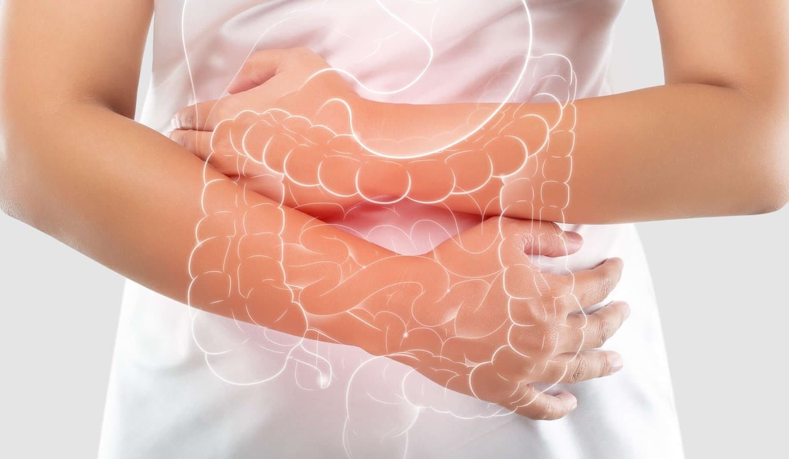
Ulcerative colitis inflames the colon and rectum, and it comes in flares.
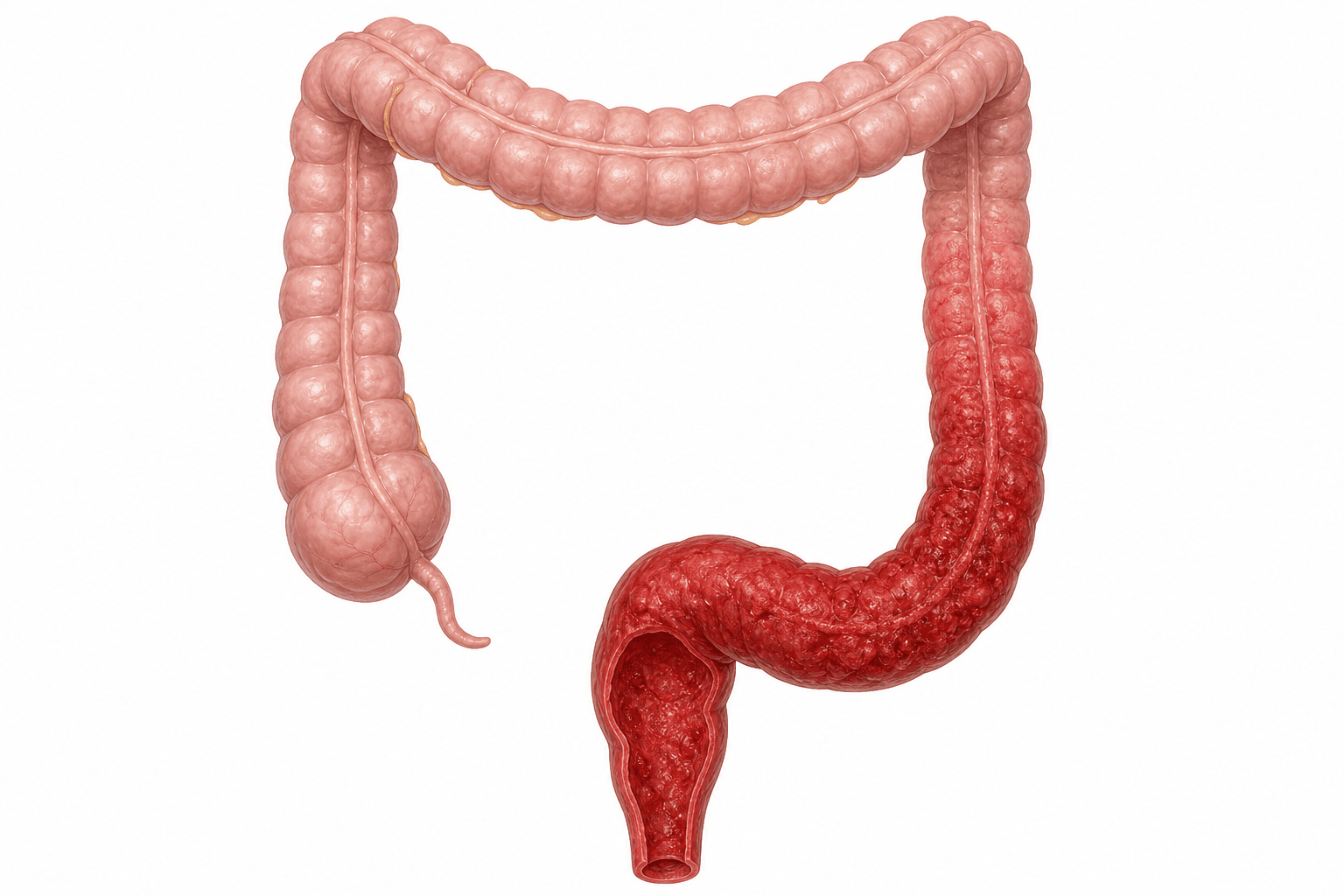
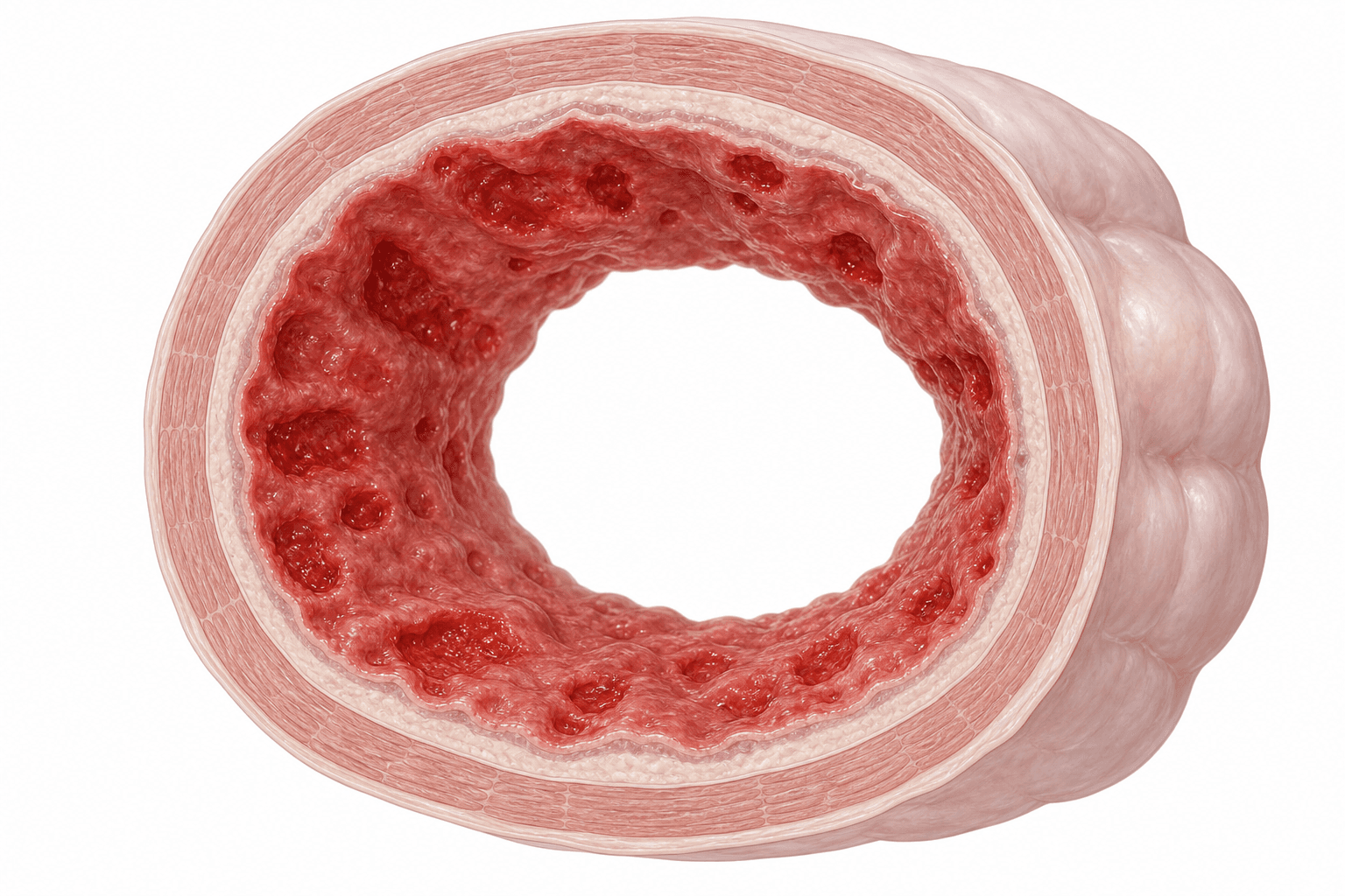
It affects the large intestine, the colon, and the rectum, causing inflammation and ulcers in the innermost lining of the digestive tract. The inflammation begins in the rectum and can extend continuously through the colon, causing tissue damage that can lead to frequent bowel movements, bleeding, and abdominal pain. Ulcerative colitis treatment is medication first, and surgery, including J-pouch surgery, for the cases medication does not control; Dr Sulaiman is the colorectal specialist patients see for that surgical side.
FRCSEd (General Surgery)Five clinics in Singapore
In short
IBD
What kind of condition it is
Ulcerative colitis is a chronic inflammatory bowel disease affecting the large intestine, the colon, and the rectum.
Rectum
Where the inflammation begins
It begins in the rectum and can extend continuously through the colon, causing tissue damage as it goes.
Flares
How it behaves over time
Patients may experience periods of active disease followed by periods of remission, so symptoms are not constant.
Under 30
When it is most often diagnosed
Ulcerative colitis can occur at any age, though it is most commonly diagnosed in people under 30.
What every patient should know
What every patient should know
Rectum
Where the inflammation begins
Source: Dr Sulaiman Bin Yusof, clinical practice
Ulcerative proctitis
The rectum only, and usually the mildest
Left-sided colitis
Rectum, sigmoid and descending colon
Pancolitis
The entire colon
Under 30
When it is most often diagnosed
Source: Dr Sulaiman Bin Yusof, clinical practice
His story
Dr Sulaiman Bin Yusof
Senior Consultant Colorectal and General Surgeon, seeing patients at five clinics in Singapore.
- MBChB (Sheffield), M.Med (Surgery), FRCSEd (General Surgery)
- Consults in English and Malay
- MOH Fellowship, Peter MacCallum Cancer Centre, Melbourne
- Singapore Health Quality Service Star Award 2023
- Dean's Honour Roll for Teaching
Symptoms
The symptoms come in flares, and these are the ones to act on.
Patients with ulcerative colitis may experience periods of active disease followed by periods of remission. These are the symptoms that most commonly bring people in, and they warrant assessment when they are new, persistent, or getting worse.
Bloody diarrhoea
The most common symptom involves loose stools mixed with blood and mucus. The frequency can range from a few to many times per day.
Abdominal pain
Patients experience cramping and discomfort, particularly in the lower abdomen. The pain often intensifies before bowel movements.
Urgency
Many patients feel a sudden, intense need to have a bowel movement. This urgency may be accompanied by an inability to hold bowel movements.
Fatigue
Chronic inflammation and possible blood loss may lead to anaemia, causing persistent tiredness and low energy levels.
Weight loss
Reduced appetite and poor absorption of nutrients can result in unintended weight loss.
Symptoms outside the gut
Some patients develop joint pain, skin problems, or eye inflammation. These symptoms occur when inflammation affects areas outside the digestive system.
These symptoms overlap with several other bowel conditions, so having one of them does not mean you have ulcerative colitis. It means the cause is worth establishing properly rather than guessing at.
Bleeding in particular has more than one possible cause, and what those are is set out in anal bleeding, causes and treatment options.
Causes and risk factors
Several factors contribute. None of them explains it alone.
Several factors contribute to the development of ulcerative colitis. These are the ones that come up most often.
Immune system dysfunction
An overactive immune system mistakenly attacks healthy cells in the digestive tract, leading to inflammation and tissue damage.
Genetic factors
A family history of ulcerative colitis increases the likelihood of developing the condition, indicating a hereditary influence.
Environmental triggers
External factors such as infections, stress, or dietary changes can initiate or worsen symptoms in individuals predisposed to the condition.
Age
Although ulcerative colitis can occur at any age, it is most commonly diagnosed in people under 30.
The three types
Which type you have is a question of how much colon is involved.
Ulcerative colitis is classified based on the extent and location of inflammation within the colon. That classification is what a colonoscopy establishes, and it is what shapes the treatment plan.
Ulcerative proctitis
Affects only the rectum, the area closest to the anus. This form tends to be the mildest, with symptoms including rectal bleeding, urgency, and tenesmus (a feeling of incomplete evacuation). Patients may achieve remission more easily compared to other types.
Left-sided colitis
Inflammation extends from the rectum up through the sigmoid and descending colon. Patients experience left-sided abdominal pain, bloody diarrhoea, and weight loss. This form affects a significant portion of the colon but may respond well to topical and oral medications.
Pancolitis
Involves the entire colon. Patients experience severe symptoms including frequent bloody diarrhoea, abdominal pain, significant weight loss, and fatigue. This form often requires more intensive medical treatment and carries a higher risk of complications.
How it is diagnosed
Confirming it, and ruling out what it resembles.
Several investigations are used to establish the diagnosis, to measure how much of the colon is affected, and to rule out other causes. No single one of them settles it alone.
Colonoscopy
A direct look at the colon and rectum
Biopsy
Tissue taken during the same procedure
Blood and stool tests
Anaemia, infection and inflammation
Imaging
CT or MRI, where complications are suspected
Colonoscopy
A thin, flexible tube with a camera examines the entire colon and rectum. The procedure allows direct visualisation of inflammation and ulcers, and enables tissue sampling for biopsy. The appearance and pattern of inflammation help distinguish ulcerative colitis from other conditions.
Biopsy
Small tissue samples taken during colonoscopy undergo microscopic examination. The analysis reveals characteristic changes in tissue architecture and inflammatory patterns specific to ulcerative colitis.
Blood tests
Blood samples check for anaemia, infection markers, and inflammatory indicators. These tests help assess disease severity and monitor treatment response, though they cannot diagnose ulcerative colitis alone.
Stool tests
Laboratory analysis of stool samples rules out infections and measures levels of inflammatory markers. These tests help differentiate between infection and disease flare-ups.
Imaging: CT and MRI
CT scans or MRIs may show bowel wall thickening or complications. These imaging techniques provide additional information about disease extent and possible complications.
What a colonoscopy involves, what the preparation is like and what the day itself looks like is on the colonoscopy page.
The condition it is most often distinguished from is the other inflammatory bowel disease, and what that involves is set out on the Crohn’s disease page.
Treatment options
From controlling the inflammation to removing the diseased bowel.
Ulcerative colitis is treated with medication that controls the inflammation, and with surgery that removes the diseased tissue. Which of them applies depends on your own case, and it is decided with you rather than for you.
Medication
Treatment often involves anti-inflammatory drugs like 5-aminosalicylates (5-ASA) to reduce bowel inflammation, immunosuppressants to control the immune response, and biologics that target specific pathways in the inflammatory process, offering effective management for moderate to severe cases.
Total proctocolectomy
This procedure involves the complete removal of the colon and rectum, effectively eliminating the diseased tissue. Waste management is tailored to the patient's condition and may involve creating an ileostomy, where waste exits the body through an external stoma into a bag, or an internal pouch constructed from the small intestine, allowing for more natural waste elimination.
J-pouch surgery
This procedure involves creating a J-shaped internal reservoir from the small intestine, allowing patients to maintain bowel continuity and function without an external bag. It is typically performed in multiple stages and is well suited to carefully selected patients who can tolerate the procedure and its recovery process.
Living with it
Between flares, most of the management is daily and unglamorous.
Managing the condition involves medication adherence, dietary adjustments, and regular monitoring. None of it replaces treatment, and together it changes how often treatment has to change.
Food diaries can help identify triggers, allowing healthcare providers to create personalised diet plans. Stress management through relaxation techniques may reduce flare-ups. Regular exercise, adequate sleep, and smoking cessation support overall health.
Routine medical check-ups ensure effective monitoring and treatment adjustments, which is why the follow-up continues once the symptoms have settled rather than stopping with them.
1 to 2 years
How often surveillance colonoscopy is advised
Source: Dr Sulaiman Bin Yusof, clinical practice
Why the monitoring continues in remission
Regular monitoring is what makes long-standing ulcerative colitis a followed condition rather than a resolved one. Remission is a good place to be, and it is not a reason to stop being watched.
What that surveillance is looking for, and how it is done, is on the colorectal cancer page.
Common questions
The questions that come up in consultation.
Can diet cure ulcerative colitis?
While diet cannot cure ulcerative colitis, certain dietary modifications may help manage symptoms. Each person responds differently to foods, making individualised dietary plans necessary.
Can ulcerative colitis cause problems outside the bowel?
Yes, patients with ulcerative colitis may have a higher risk of developing osteoporosis, liver diseases like primary sclerosing cholangitis (PSC), or blood clots. Regular monitoring helps address these risks early.
Will I need regular colonoscopies after diagnosis?
Patients with long-standing ulcerative colitis, especially pancolitis, are advised to undergo regular colonoscopies every 1 to 2 years to monitor for colorectal cancer, starting 8 to 10 years after diagnosis.
What are the treatment options, and does everyone need surgery?
Treatment often involves medication. Anti-inflammatory drugs such as 5-aminosalicylates reduce bowel inflammation, immunosuppressants control the immune response, and biologics target specific pathways in the inflammatory process for moderate to severe cases. Where surgery is used, a total proctocolectomy removes the colon and rectum completely, and waste management is then tailored to the patient's condition. Which of these applies to you depends on your own case, and Dr Sulaiman will go through that at consultation rather than leave you guessing.
If I need surgery, will I have to live with an external bag?
Not necessarily. After the colon and rectum are removed, waste management is tailored to your condition. One option is an ileostomy, where waste leaves the body through an external stoma into a bag. The other is an internal pouch constructed from the small intestine, which allows for more natural waste elimination. J-pouch surgery creates a J-shaped internal reservoir from the small intestine so that bowel continuity and function are maintained without an external bag. It is typically performed in multiple stages and is well suited to carefully selected patients who can tolerate the procedure and its recovery process.
Book a consultation
Are your symptoms affecting your quality of life? A proper assessment is where control starts.
If bloody diarrhoea, abdominal pain or urgency are affecting your quality of life, book a consultation for an assessment and a personalised treatment plan built around your own case. Dr Sulaiman sees patients at five clinic locations in Singapore, and self-referrals are welcome.